

Advanced Technology
Cone Beam CT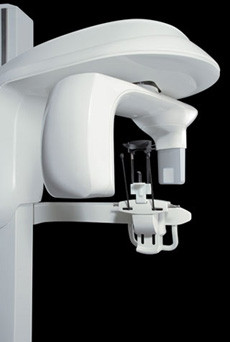
Our practice utilizes state-of-the-art, small-volume cone-beam CT (computed tomography) technology that provides highly accurate, 3-D radiographic images for the diagnosis, planning and treatment of endodontic disease. This allows three-dimensional visualization of teeth, bone, sinuses and surrounding structures with minimal radiation to the patient, enabling a level of anatomical accuracy and patient care not possible with 2-D technologies (regular dental x-rays). With the addition of cone-beam CT technology to our office, our practice is committed to providing innovative, high-quality, patient care.
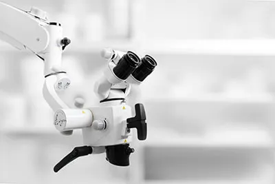 Surgical Microscopes
The introduction of the surgical microscope has revolutionized the field of Endodontic Microsurgery. We have invested in the very best quality surgical microscopes, by Carl Zeiss, that provide unparalleled magnification and illumination for our surgical procedures.
Our success depends on us being able to see the minutest of details – you cannot treat what you cannot see. In addition, we have incorporated the latest high-definition video allowing us to record our procedures with unequaled clarity and detail.
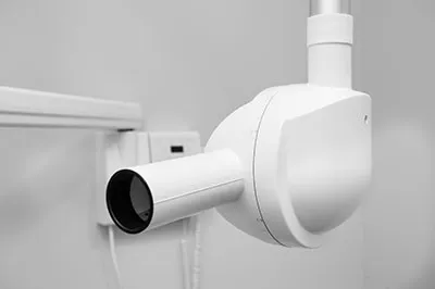 Digital Imaging
Oakmont Endodontics & Sherman Oaks Endodontics carefully chooses which and when radiographs are taken. There are many guidelines that we follow. Radiographs allow us to see everything we cannot see with our own eyes. Radiographs enable us to detect cavities in between your teeth, determine bone level, and analyze the health of your bone. We can also examine the roots and nerves of teeth, diagnose lesions such as cysts or tumors, as well as assess damage when trauma occurs.
Dental radiographs are invaluable aids in diagnosing, treating, and maintaining dental health. Exposure time for dental radiographs is extremely minimal. Oakmont Endodontics & Sherman Oaks Endodontics utilizes Digital Imaging Technologies within the office. With digital imaging, exposure time is about 90 percent less, compared to traditional radiographs. Digital imaging can also help us retrieve valuable diagnostic information. We may be able to see cavities better.
Digital imaging allows us to store patient images and enables us to quickly and easily transfer them to specialists or insurance companies.
Digital X-Rays:
Digital X-rays offer more precision since we view the image on a computer monitor, instead of holding up a 35mm film to the light. Digital X-rays result in 1/6th of the radiation exposure to you.
